St. Jude Family of Websites
Explore our cutting edge research, world-class patient care, career opportunities and more.
St. Jude Children's Research Hospital Home

- Fundraising
St. Jude Family of Websites
Explore our cutting edge research, world-class patient care, career opportunities and more.
St. Jude Children's Research Hospital Home

- Fundraising
Organic origami: Unfolding the mysteries of biomolecular structures, dynamics, and functions
The fundamental principles of origami, the Japanese art of paper folding, have endured for generations.
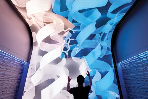
Rooted in symmetry and precision, origami involves basic folds that contribute to a larger, more complex vision. In biology, underlying almost every cellular process is a protein that must fold with precision into an intricate three-dimensional structure to carry out its function.
At St. Jude, researchers strive to understand the processes that govern biological function by teasing apart the organic origami of biomolecular activity like never before.
Capturing transporter structures paves the way for drug development
Transporters are proteins that move essential substances such as ions, neurotransmitters, and nutrients across membranes. Modulating these molecular gatekeepers can be a potent therapeutic route; thus, gaining a fundamental understanding of their intrinsic structure is paramount to accomplishing this task.

Insight from structural analysis allowed Chia-Hsueh Lee, PhD, to understand how VMAT2 transports neurotransmitters like serotonin into vesicles.
Chia-Hsueh Lee, PhD, Department of Structural Biology, studies the structures and dynamics of membrane transporters to better understand their function and links to disease. Sphingosine-1-phosphate (S1P) is an important signaling molecule that regulates the immune system, blood vessel formation, auditory function, and the integrity of epithelial and endothelial membranes. Spinster homolog 2 (Spns2) is an S1P transporter that sits on the cell membrane and moves S1P across it to be released into the extracellular space.
In a paper published in Cell, Lee and colleagues reported the six structures of Spns2 that they obtained using cryo-electron microscopy (cryo-EM), including two functionally relevant intermediate conformations (shapes) that tie together the Spns2 inward- and outward-facing states, illuminating how Spns2 functions. The findings reveal the structural basis of the S1P transport cycle and how a potential Spns2-targeted therapeutic binds. They found that the Spns2 inhibitor 16d blocks transport activity by locking Spns2 in its inward-facing state.

We hope our structural information will pave the way for the development of improved, more specific small molecules with higher potency against Spns2 in the future.
Structural Biology
In addition to Spns2, Lee also studies neuronal communication, which relies on releasing neurotransmitters from within the cell to the synaptic space between neurons. To transport neurotransmitters to the cell membrane for their release, the cell packages them into “cargo ships” called vesicles. Vesicular monoamine transporters (VMATs) are proteins located on the membranes of these vesicles and act as loading cranes to move specific neurotransmitters called monoamines from the cytoplasm into the space within the vesicle.
In work published in Nature, Lee used cryo-EM to capture multiple structures of VMAT2. “This transporter is a target for pharmacologically relevant drugs used in the treatment of hyperkinetic disorders such as chorea and Tourette syndrome,” Lee said.
Lee’s group captured this dynamic protein in multiple states and demonstrated two distinct binding modes of two drugs, reserpine and tetrabenazine, addressing a 40-year-old question regarding their binding mechanisms. Further, the researchers used a serotonin-bound structure to identify the mechanism the transporter likely uses to engage all monoamines.
Vesicles are one way of transporting molecules around the cell. Some proteins need to be transported to distant cellular regions, such as cilia, which are hair-like projections that control many signaling processes. Cilia lack the machinery to synthesize proteins, so intraflagellar transport (IFT) complexes, like IFT-A and IFT-B, form train-like structures to bring proteins into and out of cilia.
In their work published in Cell Research, Ji Sun, PhD, Department of Structural Biology, and colleagues presented the structure of the IFT-A complex and its assembled train form. The structure revealed previously unknown zinc-binding sites in IFT-A. Zinc-binding sites are important to a protein domain called zinc fingers, which are critical for certain protein−protein interactions. This explained some poorly understood connections within the train complex.
“Now that we have the structure, we can tell how IFT-A trains assemble to mediate ciliary cargo transport. We can also map mutations that disrupt this vital biological process and design clinical interventions,” said Sun.
The work serves as a foundation for studying diseases of the brain, kidney, skeleton, and eyes caused by defective cilium transport, which were previously difficult to investigate.

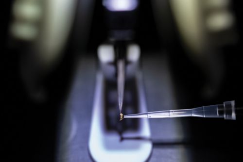
Protein structure studies in the St. Jude Cryo-Electron Microscopy Center begin with a single drop of protein in solution placed on a copper or gold grid.
Structures offer a framework for understanding how proteins work together
Proteins are not static. They move, bend, fold, and flex, offering multiple dynamic structural forms capable of different functions. Visualizing different forms of proteins during their activity cycle gives us a more complete understanding of their dynamics and how their structures relate to their function or dysfunction.
In addition to their work with IFT-A, Sun’s group reported the complex structure of two Parkinson’s disease–related proteins in Science. This was a follow-up study to previous work on leucine-rich repeat kinase 2 (LRRK2), a protein kinase that modifies other proteins through phosphorylation.
“In that first paper, we got the structure of LRRK2, but that structure showed an inactive conformation,” Sun explained.
Sometimes, it takes binding to another protein to trigger the protein’s active state. Here, the missing piece was Rab29, a member of the Rab GTPase family that regulates cellular trafficking and modulates the activity of LRRK2.
Using cryo-EM, the researchers determined the first structures of the Rab29–LRRK2 2 complex. The structures included an unexpected tetramer comprising four pairs of Rab29–LRRK2 that has been shown to assemble only at the membrane surface in cells. This tetramer finally revealed the active form of LRRK2, informing the researchers on the biological path toward protein activation and function.
“We proposed a transition from monomer to tetramer upon membrane recruitment, wherein LRRK2 becomes active,” Sun explained. “These structures provide much-needed insights for medicinal chemists to design novel inhibitors against LRRK2 for Parkinson’s disease treatment.”
LRRK2’s complex with Rab29 is an example of a protein−protein interaction. Such interactions often drive protein function, occur universally throughout all organisms, and can occur between different proteins or multiple copies of the same protein.
In research published in Molecular Cell, Tanja Mittag, PhD, Department of Structural Biology, revealed structures of speckle-type POZ protein (SPOP). SPOP is involved in identifying and breaking down other proteins the cell no longer needs and is the most frequently mutated protein in prostate cancer. When SPOP is dysregulated, it can dramatically affect protein levels, triggering disruption of cellular processes and altered signaling pathways, ultimately leading to diseases like cancer.

[SPOP mutations in prostate cancer] are well understood. However, mutations found in patients with other forms of cancer, especially endometrial cancer, were puzzling [because] the mutated sites did not seem important for SPOP function.
Structural Biology
Determining SPOP’s three-dimensional structure enabled the researchers to see how multiple copies of the protein assemble to form long chains and to identify key interactions between the assembled proteins that had not been seen before. The SPOP interaction interfaces contain many endometrial cancer–linked mutations, which may explain how SPOP contributes to disease.
Single-molecule imaging gives a new view of critical cellular structures
As Mittag’s research shows, defects in protein assembly can have disastrous results. Multiple approaches are required to reveal the mechanisms underlying protein assembly and the relation of those mechanisms to disease.
Led by Scott Blanchard, PhD, Department of Structural Biology, scientists at St. Jude and Rockefeller University combined their expertise to better understand the structural relations between the function and dysfunction of the cystic fibrosis transmembrane conductance regulator (CFTR) protein. CFTR is an anion channel, and mutations in CFTR cause cystic fibrosis, a fatal disease with no cure.
Previous CFTR studies enabled the researchers to see the channel when it is either open or closed, but the transition between those two states has been incompletely understood. By deploying single-molecule fluorescence resonance energy transfer (smFRET), combined with channel conductance measurements, the team was able to provide pivotal insights into the moving pieces of the CFTR machinery and how disease mutations and small-molecule therapies affect the protein’s function.
In Nature, Blanchard’s team reported that two nucleotide-binding domains of CFTR dimerize (combine), and this conformational change drives the channel opening. Drugs used to treat cystic fibrosis enhance channel activity by increasing dimerized channel-opening probabilities. Mutations that cause cystic fibrosis can reduce the efficiency of dimerization.

There are very few proteins that are more relevant for treating disease than CFTR because treatments for cystic fibrosis aim at ameliorating the defects in the mutant forms of this protein.
Structural Biology
In addition to using smFRET to better understand CFTR, Blanchard’s laboratory used the approach to gain a new understanding of a key biomolecular workhorse: the ribosome. After transcription occurs, the resulting messenger RNA is transported to the ribosome, where it is translated into the protein encoded by the RNA sequence. In research published in Nature, Blanchard and his team examined the human ribosome-decoding mechanism for the first time.
The researchers explored how quickly human ribosomes undergo the various decoding steps compared to the speed of comparable decoding steps in bacterial ribosomes. This work revealed kinetic and structural distinctions between the two species that shed light on regulatory mechanisms that may help guide the treatment of human diseases.
“Bacterial ribosomes have been well studied for many decades, but careful mechanistic studies have been missing on human ribosomes,” said Blanchard. “We’re very interested in human ribosomes because this system has shown potential to be targeted for clinical treatments for cancer and viral infections, and deeper knowledge of this complex molecular machine may inform new therapeutic strategies.

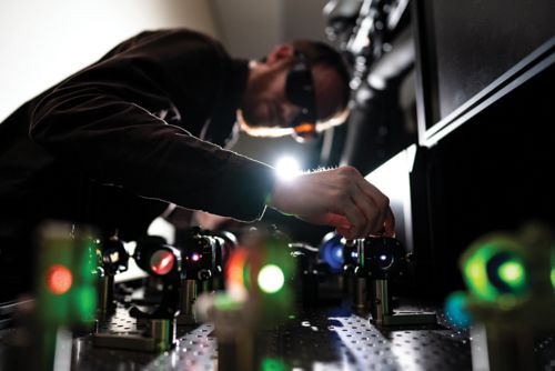
Studying protein dynamics at single-molecule resolution requires the resources and expertise of the St. Jude Single-Molecule Imaging Center.
How altered DNA structure causes cancer
Many cancers are caused by fusion oncoproteins, biomolecules that aberrantly form when a DNA rearrangement results in two proteins combining.
Many cancers are caused by fusion oncoproteins, biomolecules that aberrantly form when a DNA rearrangement results in two proteins combining. “Fusion proteins are known to be oncogenic drivers in upwards of 15% of human cancers,” said Richard Kriwacki, PhD, Department of Structural Biology.
Fusion oncoproteins can undergo a process called biomolecular condensation, wherein biomolecules separate from the surrounding environment to form their own compartments, akin to oil droplets in water. “We hypothesized that gaining the ability to form condensates could be linked with the oncogenic properties of fusion oncoproteins,” Kriwacki explained.
In a paper published in Nature Communications, the researchers revealed that 58% of the almost 200 fusion oncoproteins they examined formed condensates. They further observed that the condensate-forming fusion oncoproteins likely promote oncogenesis by altering gene regulation or cell-signaling pathways. To validate their observations, the researchers created a machine-learning algorithm using 25 recurring features of condensate-forming fusion oncoproteins. This predicted that more than 67% of the approximately 3,000 additional fusion oncoproteins tested likely form condensates, highlighting this property as a potential therapeutic vulnerability.

By grasping the underlying mechanisms, we are setting the stage for potential innovative therapeutic approaches against fusion oncoprotein–driven cancers.
Structural Biology
Biomolecular structure regulates gene expression
Protein folding is not the only biomolecular structure facet governing cellular processes. DNA also has structure regulated at multiple levels, from the chemical bonds between nucleotides that form the double helix to gene-regulating transcription factors that control access to and expression of DNA.
Myriam Labelle, PhD, Department of Oncology, has linked gene accessibility to the process of metastasis, exploring the driving forces of the movement and spread of cancerous cells through the body.
In a work published in Science Advances, Labelle reported that overexpression of the transcription factor ZBTB18 limits chromatin accessibility at gene locations important for metastasis, thereby decreasing the tumor cells’ ability to metastasize.
“Since it’s restricting chromatin accessibility, by overexpressing ZBTB18, we are essentially blocking the cells from being able to see the metastatic cues that they would normally respond to if the chromatin were more open,” explained Labelle.
These cues are tied to the cell’s plasticity, or adaptability, based on different environments. Considering that approximately 90% of cancer-related deaths stem from metastasis, Labelle’s work has the potential to motivate future therapeutic advances.
While DNA accessibility can be limited by modulating the expression of proteins like ZBTB18, proteins called pioneer transcription factors control their target DNA’s expression even within compacted chromatin. This feature is vital to kickstarting gene expression during various cellular processes.
To better understand how pioneer transcription factors access tightly wound DNA, Mario Halic, PhD, Department of Structural Biology, investigated how the pioneer transcription factor Oct4 cooperatively interacts with nucleosomes, the basic subunits of chromatin that consist of DNA tightly wound around a core of eight histone proteins, like thread on a spool. This structure not only packages DNA to fit inside the nucleus but also protects the genome from DNA-damaging perturbations. This structure is designed to safeguard the genetic material, and disruption to the factors that regulate it can lead to disease.

Building on prior work to understand the dynamic behavior of nucleosomes, we wanted to understand how other factors might utilize those dynamics to access chromatin.
Structural Biology
The researchers observed that the initial binding of an Oct4 molecule “fixes” the nucleosome in a position that increases the exposure of other binding sites, thus promoting the binding of additional transcription factors and explaining transcription factor cooperativity. They found that Oct4 contacts histones to promote chromatin decompaction. Cooperativity is further facilitated by post-translational modification of the histones, primarily the addition of acetyl groups to the histone. These findings explain how the epigenetic landscape, changes in gene expression such as post-translational modification that don’t involve alterations to DNA sequence, can regulate Oct4 activity to ensure proper cell programming.

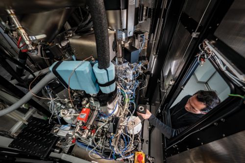
The installation of a new Krios G4 microscope allows for the investigation of biomolecules in their native environment through cryo-electron tomography.
From fundamental biology to therapeutic insights
While our understanding of biomolecular structure is the first foothold in designing new compounds for therapy, understanding how these compounds subsequently interact within the complex physiology of the body can have vast implications for therapeutics’ success or failure. For example, the protein ABCG2, responsible for removing toxins from the cell, also indiscriminately removes many kinds of chemotherapeutics, which can be a serious problem in targeting and killing cancer cells.
Research published in Nature Communications by John Schuetz, PhD, Department of Pharmacy and Pharmaceutical Sciences, addressed how ABCG2 works. ABCs are usually responsible for removing hydrophobic (water-hating) molecules, such as lipids. ABCG2 is unique because it is also involved in removing hydrophilic (water-loving) molecules, making it capable of removing many types of anti-cancer drugs from cells and limiting therapeutic efficacy.
The researchers set out to find the exact residues giving ABCG2 the capability to bind and remove hydrophilic molecules. The study highlighted the positioning of two specific amino acids in the protein’s active site, a threonine and an asparagine, the distinguishing feature of which is that they are polar and, therefore, hydrophilic. Mutating these to a featureless alanine demonstrated that the amino acids were crucial for binding hydrophilic chemicals.
ABCG2 inhibitors are often combined with chemotherapeutics, but preventing ABCG2 function can result in off-target detrimental effects. “The goal now is to design ABCG2 inhibitors that have minimal effect on normal tissues but target the tumor,” Schuetz said. This mechanistic insight can inform efforts to design more effective, less detrimental inhibitors that target the binding site threonine in ABCG2 and pair it with hydrophilic chemotherapies used in cancer treatment.
As technologies develop, understanding biomolecular structure at an atomic level has never been more critical to combating disease. The complexity of organic origami begets the wonders of the relationship between structure and function. At St. Jude, researchers will continue to uncover these wonders, one fold at a time.