St. Jude Family of Websites
Explore our cutting edge research, world-class patient care, career opportunities and more.
St. Jude Children's Research Hospital Home

- Fundraising
St. Jude Family of Websites
Explore our cutting edge research, world-class patient care, career opportunities and more.
St. Jude Children's Research Hospital Home

- Fundraising
Pediatric rhabdomyosarcoma is a cancerous tumor that arises in soft tissue, such as muscles, and consists of multiple disease subtypes. The origin of one of the most aggressive subtypes, fusion-positive rhabdomyosarcoma, has been a long-standing mystery.
Fusion-positive rhabdomyosarcoma cells express a hybrid protein (or oncoprotein) that results from the fusion of two genes, PAX3 and FOXO1. Pediatric fusion-positive rhabdomyosarcoma has a poor prognosis and is challenging to treat. Rhabdomyosarcoma cells appear muscle-like; however, fusion-positive tumors can form in areas without skeletal muscle. They also occur as diffuse tumors that resemble metastatic disease.
Scientists at St. Jude have shown in proof-of-concept models that expressing the fusion oncoprotein in either muscle or blood vessel (endothelial) cells produces aggressive tumors, which are molecularly identical to fusion-positive rhabdomyosarcoma. The researchers generated models to find where and how these fusion-positive tumors arise. Their findings, published in Nature Communications, have implications for treating aggressive rhabdomyosarcoma.

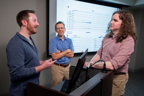
Co-first author Randolph Larsen IV, corresponding author Mark Hatley, MD, PhD, both of the Department of Oncology, and co-author Brian Abraham, PhD, Department of Computational Biology, discuss their paper.
“When we compared the tumors that develop from muscle or endothelial cells, they were nearly indistinguishable,” said corresponding author Mark Hatley, MD, PhD, Department of Oncology. “We performed detailed analyses, including looking at gene expression, chromatin architecture, and enhancer accessibility. Despite how hard we looked, we could not find a single distinguishing feature that told us that the tumor from the endothelial cells came from endothelial progenitors and not muscle cells.”
Hatley’s lab generated a mouse model expressing the PAX3–FOXO1 fusion oncoprotein in blood vessel cells. The researchers compared this new model to a previously established mouse model of fusion-positive rhabdomyosarcoma that arises from muscle cells. The study indicates that it may not be possible to find the cellular origin of these cancerous cells using standard methods. Instead, focusing on the fusion oncoprotein’s effects may be a better way to identify potential therapeutic interventions.
To explore this alternative approach, the Hatley lab developed a model to examine the genetic and epigenetic effects of the fusion oncoprotein in human cells more closely. They exploited the oncoprotein’s strength as a cancer driver in human induced pluripotent stem cells (hiPSCs), which can differentiate into any cell type. The scientists first differentiated the hiPSCs into endothelial cells and then added the fusion oncoprotein. This in vitro system enabled the team to look even closer at what happens when the cells undergo malignant transformation.
The fusion oncoprotein still acted as a potent activator of muscle cell fate in the system. The aberrant protein bound to DNA regions that enhance the expression of genes well known to be major regulators of muscle cell development, such as MYOD1. These findings serve as a proof of concept that this hiPSC system can be used to find downstream targets of the PAX3–FOXO1 oncoprotein and increase confidence in any targets found in future studies.