St. Jude Family of Websites
Explore our cutting edge research, world-class patient care, career opportunities and more.
St. Jude Children's Research Hospital Home

- Fundraising
St. Jude Family of Websites
Explore our cutting edge research, world-class patient care, career opportunities and more.
St. Jude Children's Research Hospital Home

- Fundraising
Your fatty acid delivery is on the way: studying cellular logistics provides disease insights

Scientist Xiao Li, Principal Investigator Chi-Lun Chang, and Scientist Rico Gamuyo from the St. Jude Department of Cell and Molecular Biology developed a fluorescence-based toolkit called FABCON.
Throughout history, cellular biology research has focused on describing different types of cells, cellular components, and how they interact and function. Chi-Lun Chang, PhD, St. Jude Department of Cell & Molecular Biology, approaches cell biology from a different perspective: logistics.
Logistics involves managing the supply and transportation of goods to where they are needed. “We face logistical issues every day,” Chang said. “How will we get to work? How will we get our groceries? Our cells and their various components face issues of logistics too.”
The human cell is a wonderfully compartmentalized system. The cell nucleus (an organelle) stores the cell’s DNA; lipid droplets (also organelles) store fatty acids and the fundamental energy molecule ATP is produced in mitochondria (yet another organelle) before it moves to the cytoplasm for use.
However, a compartmentalized system produces logistical concerns. Imagine a bustling city where goods must move quickly and efficiently between businesses. Many issues arise from delays in material delivery or loss of packages in transit. These issues are not only inconvenient but can grind construction projects, business activities, and daily life to a halt. Similarly, raw materials, energy and signaling molecules must also travel quickly and efficiently between different cellular organelles with different specialized functions.
Chang studies how human cells handle their logistics. It is a fundamental process, but there is still much to learn. As described in a recent Journal of Cell Biology, Chang’s lab developed a fluorescence-based toolkit called FABCON (Fluorescent And Bioluminescent Contact Observation Nexus), to detect nanometer-scale contact sites where organelles such as lipid droplets communicate and exchange material with other organelles inside cells. With tools such as FABCON, researchers can study how logistical errors inside human cells can lead to various diseases, including neurological conditions such as dementia.
How do lipid droplets efficiently deliver fatty acids?
Chang is particularly interested in how fatty acids move inside the human cell. He is studying lipid droplets, specialized organelles that store fatty acids. These droplets are tiny in many cell types, but in professional fat-storing cells can be quite large.
“You can think of lipid droplets as warehouses of fatty acids, an essential material for your cells,” Chang said. “Fatty acids are the building blocks of the lipids that form all cell membranes. They are also signaling molecule and a high-energy fuel your cells can burn via your mitochondria.”
Fatty acids are so essential that they are kept in surplus since so many different organelles need quick access to them.

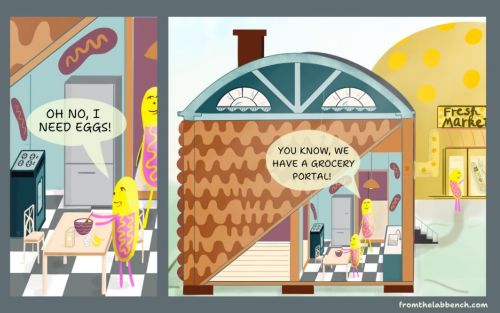
Imagine the organelle contact site as a “portal” to a grocery store. On the left, a character in a house resembling a mitochondrion is cooking but realizes they forgot to buy eggs. On the right, another character explains, “You know, we have a grocery store portal!” A tunnel is sending materials directly from the market to the house’s kitchen. This is an analogy for how organelles deal with the logistics of exchanging resources through contact sites.
Access to energy is a critical logistics issue in many different systems. Proximity to food motivated the invention of home refrigerators and food delivery services. But in the microscopic universe of the human cell, the few feet you need to walk to get to your refrigerator for a snack might take weeks for a molecule like a fatty acid to travel. So, how do lipid droplets efficiently deliver fatty acids to other organelles that process them?
You can often find Chang peering through a microscope in a darkened room at images of brightly colored components inside cells. He’s looking for where and when different cellular organelles, such as mitochondria and lipid droplets, come into extremely close contact. While we humans have developed various technologies to help us with our logistics — conveyor belts, geolocation technologies for tracking, delivery services, computers, and 3D printing — cells have implemented an innovation called the contact site.
At contact sites, organelles are as little as 20 nanometers apart, a gap 5,000 times smaller than the width of a human hair and shorter than the wavelength of visible light. These sites play a crucial role in cellular logistics, facilitating the rapid exchange of materials between organelles. Through contact sites with lipid droplets, mitochondria can quickly access fatty acids during intense energy use.
Chang and his lab are interested in studying contact site dynamics and how they are regulated, when they form, and for how long. Because of technical limitations, researchers have not been able to answer these questions until now.
How do you monitor contact sites smaller than the wavelength of light?
Contact sites between organelles are smaller than the smallest objects that can be directly observed using light microscopy. In the 1950s, researchers first observed organelle contact sites using electron microscopy, reflecting electrons off a surface instead of the photons of conventional light microscopy. The wavelength of high-energy electrons is thousands of times smaller than the wavelength of visible light, making electron microscopy better for observing nanometer-sized objects.
There’s only one problem: cells must be heavily processed and embedded in a thin slice of plastic for electron microscopy. In other words, the cells are no longer alive, making it impossible to observe dynamic processes such as the active formation of contact sites.
In the last decade, researchers have devised an elegant solution for observing interactions between tiny components inside living cells with light microscopy. They’ve taken fluorescent proteins, such as green fluorescent protein (GFP), and split them in half. Neither half fluoresces on its own, but when the halves come close enough together, they light up under a microscope. Researchers originally developed this “split fluorescence reporter” system to detect protein-protein interactions, attaching the two halves of a fluorescent reporter to two proteins of interest. Fluorescence indicates the proteins are interacting.
“You can use this same technique for detecting organelle-organelle interactions,” Chang said. “But there are some major issues.”
GFP, the most common split fluorescence reporter for cell research, has an irreversible interaction. When the two halves of the split reporter come into contact, they lock in space and stay together. Imagine that every time you tried to track a delivery, a subscription service was created that delivered the same package to your doorstep daily. That would be inconvenient. Similarly, you can’t track the dynamic and transient process of organelles making contact if your reporter system makes them lock together when they come into contact.
Chang and his colleagues had an idea to overcome this challenge and study the dynamic process of lipid droplet contact sites with mitochondria and other organelles: they needed a reversible split fluorescence reporter that would detect but not artificially interfere with or “lock in” contact sites. They also needed to develop a way to target one-half of this reporter to lipid droplets and not any other organelles.
A fluorescence toolkit for studying lipid droplet dynamics
Soon after Chang set up his lab at St. Jude, he heard a collaborator had developed a new reversible fluorescence reporter called splitFAST. Chang’s lab got to work on adapting it for lipid droplet research.
It took three years, but Chang’s lab ultimately developed the FABCON toolkit, which involves a splitFAST reporter that targets lipid droplets and can detect their interactions with mitochondria and other organelles.
Chang’s group overcame two significant challenges in developing this toolkit. First, they incorporated a repeat of a low-affinity motif that strongly targets lipid droplets, which produces a strong “on-off” signal to detect contact sites and lowers background noise. Second, the group ensured the split reporter system did not interfere with naturally occurring contact sites.
Chang and his lab have now used FABCON to detect small changes in lipid droplet contact sites allowing them to study contact dynamics between lipid droplets and other organelles.
In the Journal of Cell Biology paper, the St. Jude researchers found that lipid droplet contact sites with the endoplasmic reticulum emerge in cells when new lipids need to be synthesized or in states of cell biogenesis. They’ve also found that transient contact sites between lipid droplets and mitochondria appear when a cell needs alternative energy sources such as fatty acids.
“This toolkit allows us to not only detect the presence of contact sites in real-time but even measure their size and understand their dynamics and regulation,” Chang said. “This is a critical ability for studying diseases like Parkinson’s disease where these contact sites may be disrupted.”
FABCON illuminates fabulous organelle contact sites
Observing organelle contact sites in action will open new avenues for disease research and give researchers unprecedented insight into cellular processes.
“The development of FABCON was a highly collaborative effort between our lab, collaborators, and core research facilities at St. Jude,” said Xiao Li, PhD, first author of the paper and a researcher in Chang’s lab.
With new research tools, investigators can ask questions that were previously unanswerable. The team aims to refine FABCON further, potentially benchmarking the affinity between different organelles to gain a deeper understanding of cellular logistics. They also hope to apply this toolkit to study a broader range of diseases, providing valuable insights that could lead to new therapeutic approaches.
“Before I worked on this project with Dr. Chang, I’d never really thought about how tiny sites of contact between two organelles could be so important to the transfer of material and cellular signaling,” said Li. “I think we’re just scratching the surface of what we can learn about cellular communication. FABCON is a powerful new tool.”


