St. Jude Family of Websites
Explore our cutting edge research, world-class patient care, career opportunities and more.
St. Jude Children's Research Hospital Home

- Fundraising
St. Jude Family of Websites
Explore our cutting edge research, world-class patient care, career opportunities and more.
St. Jude Children's Research Hospital Home

- Fundraising
Chi-Lun Chang Lab
Investigating the dynamic spatial organization of inter-organelle logistics
About the Chang Lab
To support the health and function of an organism, nutrients must be converted into usable products via specific and localized processes. There is a sophisticated logistic network within individual cells that ensures proper distribution and trafficking of these nutrients and their byproducts. Our lab is interested in understanding how this logistics network is established and maintained. We focus on two fundamental aspects of biology: energy homeostasis and protein secretion.

Our research summary
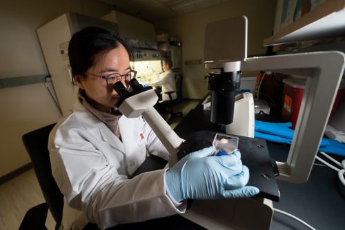
Nutrients and metabolites need to be distributed carefully between organelles within cells to integrate cellular function and meet metabolic demands in a human body. Our laboratory investigates the formation, organization, regulation, and function of a complex inter-organelle logistic network. We address these questions via cutting edge light and electron microscopy tools in conjunction with a multidisciplinary approach, including molecular and cell biology, biochemistry, in vitro reconstitution, real-time metabolic analyses, and genome-editing technology. Our goal is to gain mechanistic insights into this logistic network in normal cells so we can translate this knowledge into the understanding of pathogenesis of diseases.
Understanding the formation and regulation of lipid droplet-organelle contact sites
Fatty acids are indispensable materials for lipid synthesis and energy production. Lipid droplets play important roles in fatty acid metabolism via functional alliances with many other organelles at contact sites. However, the molecular basis of lipid droplet-organelle contact sites formation and regulation are largely unknown. This is due, in part, to the lack of tools that can visualize such dynamic nano-architecture, as well as the scarcity of known tethering components. Our team plans to engineer synthetic tools that selectively label lipid droplet-organelle contact sites with minimal perturbation to cells. These tools will enable us to quantitatively describe the dynamics of contact sites in conditions of energy surplus or starvation. We also aim to identify and characterize tethering components by mapping the proteome at these sites using proximity labeling enzymes and by CRISPR-based knockout screens..

Deciphering how the tubulin code collectively regulates organelle distribution involved in energy homeostasis
It is yet unknown how cells coordinate the proximity of organelles to form contact sites in the first place. Evidence suggests that tubulin codes, generated by posttranslational modifications, create identifying landmarks. We hypothesize that the tubulin code dictates the location and duration of lipid droplet-organelle contact sites to regulate trafficking energy homeostasis. We are addressing this question from the perspectives of code-writing enzymes and code-recognizing effectors. We are applying gene editing technology to modulate coding/recognition machinery and analyze distribution, architecture, and function of these contact sites. Aberrant regulation of the tubulin code are associated with neurodegenerative diseases and defective energy homeostasis. Our work stands to provide a new perspective on the pathogenesis of neuron cell dysfunction as it relates to energy homeostasis at lipid droplet-organelle contact sites.
Investigating the formation and function of early secretory compartments
A conserved secretory pathway is responsible for trafficking membrane and secretory proteins made in the endoplasmic reticulum (ER). The first step in this pathway is to sort and concentrate protein cargos into ER exit sites (ERESs). Our previous work revealed a dynamic, complex tubule network of early secretory compartments in intact mammalian cells and provided an updated conceptual framework for understanding how the secretory pathway functions and adapts to deliver diverse cargo types to the Golgi. We are now interested in understanding how these membrane organizations are formed. Our lab is developing synthetic degradation systems and in vitro reconstitution assays to identify critical mediators and recapitulate ERES formation. Ultimately, we aim to understand how this early secretory pathway functions and adapts to accommodate metabolic demands.
Selected publications
Contact us
Chi-Lun Chang, PhD
Assistant Member, Cell and Molecular Biology
MS 340, Room M4426
St. Jude Children's Research Hospital
Follow us

Memphis, TN, 38105-3678 USA GET DIRECTIONS