St. Jude Family of Websites
Explore our cutting edge research, world-class patient care, career opportunities and more.
St. Jude Children's Research Hospital Home

- Fundraising
St. Jude Family of Websites
Explore our cutting edge research, world-class patient care, career opportunities and more.
St. Jude Children's Research Hospital Home

- Fundraising
Bone marrow transplantation for sickle cell disease improves myocardial fibrosis

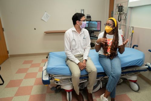
Akshay Sharma, MBBS, Department of Bone Marrow Transplantation & Cellular Therapy talks with St. Jude patient Nia Mullins in the clinic.
Of all the sounds you hear as you walk the halls of a hospital, the most iconic is the rhythmic beeping of a heart monitor. The heart is so synonymous with healthcare because it is among our most vital organs – it’s function or dysfunction being the difference between life and death. Preserving heart health is important for all people, but more so for individuals living with sickle cell disease, whose condition damages the heart muscle.
Sickle cell is an inherited trait which causes red blood cells to form a crescent or sickle shape. These sickled cells stick together, clogging blood vessels and causing pain and a host of other symptoms that can last a lifetime. People with sickle cell disease are born with the condition, and the damage it causes accumulates over time, making it critically important to understand the impact of the disease on organs such as the heart – and how to mitigate any damage.
Until now, there has been no known way to reverse heart muscle damage in patients with sickle cell disease. Even if patients can undergo bone marrow transplantation, the current standard curative therapy for sickle cell disease – it was long held that any damage caused by the disease would remain. However, new results from investigators at St. Jude Children’s Research Hospital and LeBonheur Children’s Hospital suggest that reversing heart damage in patients with sickle cell disease is possible.
“Sickle cell disease is like a hammer which keeps chipping away at the brick wall of your health,” said first and corresponding author of the new study, Akshay Sharma, MBBS, MSc, St. Jude Department of Bone Marrow Transplantation & Cellular Therapy. “The hammer hits the wall every day, chipping one brick at a time. When we do a bone marrow transplant, we take the hammer away, but the wall is still damaged.”
“One of the questions that we wanted to ask was what happens to the heart muscle after transplant – and to be honest with you, we didn’t expect things to improve,” added Sharma. “But they do.”
Understanding the cardiac impact of sickle cell disease
Doctors have known for many years that individuals with sickle cell disease have a higher risk of developing cardiac complications than those who do not have sickle cell disease. Patients with sickle cell disease can have arrhythmias (think of them like electrical issues of the heart), enlarged hearts, and damage to the heart muscle in the form of myocardial fibrosis.
When injuries, like falling onto our knees, heal, they may leave behind a scar. Fibrosis is like that scar, but while scarring is a normal part of healing, if it occurs repeatedly your entire life, it can cause a problem. If you keep falling on your knee every day, week after week, that scar will become very large, restricting function so that you cannot bend it or move as freely as you once did. In sickle cell disease, the heart muscle can be subject to continuous damage over time causing a buildup of fibrosis of the heart. Myocardial fibrosis may be a contributing factor in cardiac arrythmias and thus sudden cardiac death.
This makes preventing cardiac complications an important goal of any sickle cell disease treatment. Preventing heart damage and improving or reversing heart damage are two very different challenges, however. Initial evidence into the effect of sickle cell disease treatment on the heart did not look promising for improving long-term damage.
?wid=500)
Sickle cell disease can cause heart problems, including myocardial fibrosis.
Cardiac MRI emerges as a gold standard to look at heart muscle
In 2023, in Blood, Sharma and colleagues including Jane Hankins, MD, St. Jude Global Hematology Program director and Department of Global Pediatric Medicine member and Jason Johnson, MD, MHS, a pediatric cardiologist and cardiac imaging expert at LeBonheur Children’s Hospital and University of Tennessee Health Science Center, showed that disease-modifying therapies such as hydroxyurea and blood transfusions do not prevent the development of myocardial fibrosis. This is true even if the therapies are started as early as 3 years of age.
But while that finding was disappointing, the study did highlight the important role that cardiac magnetic resonance imaging (MRI) can play in understanding how sickle cell disease changes the heart muscle.
The study showed that cardiac MRI is sensitive enough to catch fibrotic changes early in life and can allow physicians to monitor changes to the heart while studying the impact of treatments for sickle cell disease. Cardiac MRI is the only current way to measure both heart muscle structure and function, unlike echocardiograms, which are restricted to measuring function.
"Cardiac MRI is the gold standard to measure cardiac ventricular volume and mass, superior to any other imaging technique,” explained Johnson. “Cardiac MRI has the added benefit of myocardial tissue characterization to identify the presence of fibrosis, edema, or inflammation that is important to define in sickle cell patients before and after bone marrow transplantation."
Through a clinical trial, SCDHCT, which was led by Sharma at St. Jude, the researchers studied the long-term impact to the heart after transplantation. In their new paper, which was also published in Blood, Sharma, Johnson and their colleagues used cardiac MRI to study the heart in patients enrolled in SCDHCT.
Transplantation can improve heart function as well as reduce fibrosis
Sharma says he expected to see that younger patients had less cardiac damage at the time of their transplant, while older patients had more, and importantly, he expected the amount of heart damage they had to stay the same after transplantation. But it didn’t.
“When we looked at the heart with cardiac MRI, we saw a gradual and definitive improvement in the amount of fibrosis in the heart,” said Sharma. “We thought that the function of the heart would improve after transplantation, and it did - but what was surprising was that the amount of fibrosis in the heart also improved over time.”
Having cardiac MRI in their toolkit enabled the researchers to make this observation. "Using cardiac MRI, we found that there was successful myocardial remodeling, defined by decreased ventricular volumes and mass with preserved systolic function, after bone marrow transplantation for sickle cell disease,” said Johnson. “Even more exciting was the improved myocardial fibrosis measured by extracellular volume, a technique only available by cardiac MRI, in these patients."
This is the first study to demonstrate that bone marrow transplantation can reduce myocardial fibrosis - something no other treatment for sickle cell disease has ever been shown to accomplish.
“Now, we have a treatment that not only prevents damage to the brick wall but can also repair the wall,” said Sharma.
While the researchers have so far only looked at the impact of transplantation on the heart, they are hopeful that other organs known to be damaged by sickle cell disease, such as the kidneys, lungs and brain, among others, might also show similar results.
“This is hopeful news for patients,” said Sharma.


