St. Jude Family of Websites
Explore our cutting edge research, world-class patient care, career opportunities and more.
St. Jude Children's Research Hospital Home

- Fundraising
St. Jude Family of Websites
Explore our cutting edge research, world-class patient care, career opportunities and more.
St. Jude Children's Research Hospital Home

- Fundraising
A glimpse beyond life and death: Exploring the biology of the MICOS complex

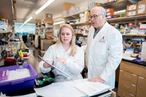
First author Stephanie Rockfield, PhD, and corresponding author Joseph Opferman, PhD, St. Jude Department of Cell & Molecular Biology, characterized a new mouse model which offers insight into the importance of MIC60, a key part of the complex responsible for mitochondrial structure and organization.
In 1945, “Mike the Headless Chicken” became a scientific legend by surviving 18 months despite missing most of his name-sake body part. Mike became a case study in neuroscience and a celebration of the brainstem’s capacity, the preservation of which allowed the chicken not just to survive but thrive. Important neurological lessons were learned from this case study, such as how structure determines function: The brainstem, known to be vital for survival, remained intact; therefore, Mike survived.
Many biological pathways are exceedingly difficult to study because disrupting them is lethal. As Mike’s case illustrates, the brain stem is vital for life, which makes studying it delicate. Breaking through this barrier to give scientists a glimpse into the biochemistry underlying such systems is a key step to developing a clearer understanding of the biological impact of these pathways.
Results from a recent Life Science Alliance publication on an essential gene in mitochondrial structure from the lab of Joseph Opferman, PhD, St. Jude Department of Cell & Molecular Biology, expounds on one component on the list of what the body absolutely cannot do without. “Immt deletion is lethal.”
Peeling back the layers of MIC60 function
Immt encodes the protein MIC60, a vital component of mitochondria. MIC60 functions within the mitochondrial contact site and cristae organizing system (MICOS). Yeast and cell culture models have identified MIC60 interaction partners and demonstrated their role in regulating cristae junction formation. However, mammalian studies are lacking, primarily demonstrating lethality upon MIC60 removal. This is not surprising, malfunctioning mitochondria essentially lead to a malfunction in every cellular process.
“Mitochondria are critical agents of biology in energy production, regulation of cell death and cellular biomass,” said Opferman. “Understanding the impact of the MICOS in mammals, which had originally been identified in yeast, intrigued us.”
Mitochondria have layers which govern function. There is an outer membrane that functions as a barrier and a specialized inner membrane with folds called cristae which increase surface area and, thus, increase capacity for energy production.
“The MICOS complex is a key player in the folding of the inner mitochondrial membrane to form cristae,” explained first author of the study, Stephanie Rockfield, PhD. “If you have aberrations in the MICOS complex, then you don’t have proper cristae architecture, and the mitochondria don’t function properly.”
While studies have shown a link between disruption of the MICOS complex and neurodegenerative disease, disruption of the gene is largely deadly. “You don’t see too many patients that have issues with the MICOS complex. They’re very rare diseases,” said Rockfield. “For MIC60, only two patients have been identified as having a possibly causal mutation in the gene, leading to developmental encephalopathy. It demonstrates what we show in our paper and what others have shown, too. If you lose MIC60, it’s embryonically lethal.”
That lethality makes studying the exact purpose of MIC60 difficult, as models don’t grow or live long enough to test.
New model offers brief glance into deadly cellular state
To address the lethality challenge, the researchers developed a mouse model that offers a vital window into the biology surrounding MIC60. This model harbors genetic markers known as LoxP sites carefully placed within the mouse genome at the location of the Immt gene. LoxP sites act as guidelines for an on-call demolition team, a role played by the enzyme Cre recombinase. Once given the signal, which, in this case, is the addition of the chemical tamoxifen, the demolition team gets to work removing the gene between the LoxP sites.
“The presence of tamoxifen triggers the expression of Cre recombinase, which will then cut out the region between the LoxP sites, leading to the end of MIC60 protein expression,” explained Rockfield. “The power of this is that it’s an inducible system, so we can assess what happens when you lose MIC60 and the MICOS complex in adult mice.”
Using the inducible model, the team pinpointed phenotypes associated with Immt deletion in healthy mice. They first observed bone marrow hypocellularity —a lower than expected number of cells.
“We initially weren’t sure whether it was bone marrow failure that was contributing to death or if it was some other tissue,” said Opferman. “We put bone marrow back in to see if it could rescue their survival. When that didn’t work, we knew something more significant was going on. That’s what led us down to the intestine.”
The scientists observed disrupted mitochondrial organization in the small intestine and colon. Mitochondria appeared significantly larger with misfolded cristae. This led to ill-functioning organs and paralytic ileus, a form of functional motor paralysis of the digestive tract.
Organ- and cell-specific gene deletion represents way forward
Intriguingly, MIC60’s significant impact on the intestinal tract may simply be tied to how long the protein hangs around. The half-life of the protein, a measure of how long it takes for half of the biomolecule to break down, is quite long — around eight days. Intestinal cells, however, turn over quite rapidly, every four to five days, meaning once the supply line for MIC60 is cut off, the intestine may just be the first to clear out lingering MIC60 and subsequently feel the hit.
Like testing a circuit breaker to find a faulty wire, Opferman hopes the model can be used to interrogate discrete organs to study what happens when MIC60 is cut off from each organ individually, even getting down to the effect on individual cells. “We want to use cell imaging to study the different domains of the MICOS complex and how they contribute to maintaining the normal structure and affect the physiology of the cell.”
The model also offers the opportunity for researchers in the field to examine MIC60 disruption in the brain. In addition to the protein’s link to developmental encephalopathy, rare heterogenous mutations (passed on from only one parent) that affect MIC60 localization to the mitochondria have been associated with hereditary Parkinson’s disease.
“Neurodegenerative models often have problems in energy production. The question is why mitochondrial abnormalities often cause these types of problems,” said Opferman.
While Mike the Headless Chicken contributed essential knowledge about the brainstem to neuroscience, the Opferman lab’s MIC60 model provides knowledge about the importance of parts of the MICOS complex. The model will act as a valuable tool to continue to explore the details of mitochondria biology in an unprecedented way.
“It sets us up where we can interrogate the structure and function of all these MICOS family members much more intently,” said Opferman. “That’s what we are pursuing right now to demonstrate the utility of what we’ve generated.”


