St. Jude Family of Websites
Explore our cutting edge research, world-class patient care, career opportunities and more.
St. Jude Children's Research Hospital Home

- Fundraising
St. Jude Family of Websites
Explore our cutting edge research, world-class patient care, career opportunities and more.
St. Jude Children's Research Hospital Home

- Fundraising
Cell and Tissue Imaging Center
A fully staffed microscopy core facility available to all St. Jude researchers
OVERVIEW
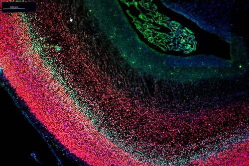
The Cell and Tissue Imaging Center is a fully staffed microscopy core facility available to all St. Jude researchers. Its mission is to provide offers access to a broad spectrum of sophisticated technologies for light microscopy, electron microscopy and image analysis. Expert professional staff are on hand to assist throughout the experimental process up to the point of publication. As well as operating instruments, helping with sample preparation, and performing analyses, staff can help select the best imaging strategies to answer specific research questions. In-depth training is also offered. The Light Microscopy and Electron Microscopy divisions within the center collaborate on correlated imaging for projects that require individual samples to be imaged using both techniques. Costs are heavily subsidized by the institution to speed discovery.

CTIC by the numbers
175+
175+ hours of LM instrument usage weekly
CTIC by the numbers
>55
More than 55 collaborative partnerships with St. Jude laboratories
CTIC by the numbers
900+
900+ samples processed for ultrastructural imaging annually

Researchers at other institutions may have similar instrumentation, but they don’t have access to a backbone of imaging scientists. We can take that cutting edge science and make it a bit cooler, and hopefully, get more data at a faster pace.
Aaron Pitre, Sr. Scientist
CTIC-Light Microscopy

IMPACT
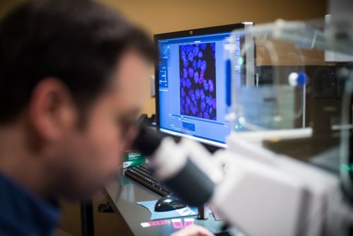
The goal of CTIC-LM is to drive and accelerate the use of advanced imaging technologies at St. Jude. CTIC-LM is staffed by PhD level imaging scientists who have years of specialized training in optics and microscopy. Due to the minimal recharge requirements, instrument rates are low, and staff have time to contribute intellectually to many projects. In the most recent quarter, CTIC-LM has engaged 85 users from 39 labs for 175 hour of instrument usage per week. It has been acknowledged for intellectual contribution on 16 peer-reviewed publications in the past year.
Available technologies
- Confocal microscopy, point-scanning and spinning disk (CLSM, SDC)
- Fluorescence correlation spectroscopy (FCS)
- Fluorescence recovery after photobleaching (FRAP)
- Fluorescence resonance energy transfer (FRET)
- Super resolution techniques including:
- Photoactivated Light Microscopy (PALM)
- Stochastic optical reconstruction microscopy (STORM)
- Structured Illumination Microscopy (SIM)
- Photoactivated Light Microscopy (PALM)
- Multiphoton Imaging (MPE)
- Selective Plane Illumination Microscopy (SPIM)
- Total Internal Reflection Fluorescence Microscopy (TIRF)
- Single Molecule Localization Microscopy (SMLM)
- Direct stochastic optical reconstruction microscopy (dSTORM)
- Point Accumulation for Imaging in Nanoscale Topography (PAINT)
- Frequency domain fluorescence lifetime imaging (FLIM)
- Live cell imaging and time-lapse imaging
- Single molecule imaging and ion imaging
- Image analysis and processing, including 3D display and deconvolution
- Slide Scanning (brightfield and epifluorescence)
- Wide-field Microscopy (transmitted and epifluorescence)
Instrumentation
- Zeiss LSM 980 AiryScan2
- Zeiss LSM 780 NLO
- Leica SP-8 with FALCON
- Zeiss lattice light sheet 7
- Nikon C2
- Marianas 1: Zeiss Axio Observer, CSU-X spinning disc, 4 diode lasers
- Marianas 2: Zeiss Axio Observer, CSU-W spinning disc, 6 diode lasers
- Zeiss Elyra PS.1
- Lightsheet Z.1
- Nikon Eclipse Ni Widefield Microscope
- AxioScan Z.1 Whole Slide Scanner
- Analysis Workstations
CTIC-Electron Microscopy

IMPACT
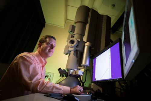
The CTIC-EM is a highly specialized resource offering full service for routine and advanced (immunolabeling, large area, and 3D imaging) electron microscopy techniques, individual consultations on project set-up, optimization, and initiation and guidance on image analysis as requested and in conjunction with the Center for Bioimage Informatics. The CTIC-EM facility collaborates with 15-20 labs at St. Jude on an annual basis and delivers ultrastructural imaging on 900 to 1000 samples each year. On average, the CTIC-EM is acknowledged on 30 peer-reviewed publications annually. Thanks to strong institutional support, CTIC-EM provides equitable access to advanced imaging equipment to all faculty at low cost, regardless of rank or experience.
Instrumentation and available technologies
- Electron Microscopes:
- Thermo Fisher Scientific Tecnai G² F20-TWIN transmission electron microscope with a Field Emission Gun and maximum operating voltage up to 200 KV and equipped for cellular electron tomography
- Thermo Fisher Scientific Helios NanoLab G3 DualBeam electron microscope with Oxford EDS detector, MAPS software, Auto Slice & View, EDS3, and monochromator, with an accelerating voltage range of 350 eV to 30 keV
- Zeiss GeminiSEM 460 field emission scanning electron microscope equipped for high-speed large area image acquisition using backscatter or STEM detectors and ATLAS AT software as well as variable pressure operation with an accelerating voltage of 20 eV to 30 keV with a maximum probe current of 40nA.
- Preparatory Equipment
- Leica Electron Microscopy Tissue Processor
- Leica UltraCut 7 and ARTOS ultramicrotomes
- Leica Automated Freeze Substitution Units and Processors
- Leica and Denton Sputter and E-beam coaters
- A workstation is available for image analysis and 3D reconstruction
Directors

Aaron Taylor
Director, Research Operations – CITC-LM
aaron.taylor@stjude.org

Cam Robinson, PhD
Director, Research Operations – CITC-EM
Cam.robinson@stjude.org