St. Jude Family of Websites
Explore our cutting edge research, world-class patient care, career opportunities and more.
St. Jude Children's Research Hospital Home

- Fundraising
St. Jude Family of Websites
Explore our cutting edge research, world-class patient care, career opportunities and more.
St. Jude Children's Research Hospital Home

- Fundraising
The “tall tail” behind ultrafast endocytosis

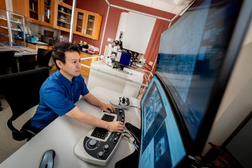
Neurons rely on ultrafast endocytosis to move molecules from outside the cell to their destination inside, a process studied by Yuuta Imoto, PhD, St. Jude Department of Developmental Neurobiology.
There are universal processes that occur across all cell types. These include glycolysis to provide energy, DNA transcription to form RNA, and RNA translation to produce a protein. Endocytosis is a universal process in almost every cell type in the human body. It can be thought of as a form of cargo transport or shipping. To move molecules from outside the cell to their destination inside, cells internalize part of their membrane and the external molecules, packing them into small shipping containers called vesicles. These vesicles then move the molecules where they need to go. After vesicles reach the destination for their cargo, they reform with another membrane, opening the vesicle and releasing the cargo molecules.
What differentiates one cell from another regarding these processes is the speed or frequency with which they occur. While all cells use endocytosis, neurons have the process down to a fine art. The rapid turnover of neurotransmitters during synaptic activity means that synaptic vesicles loaded with neurotransmitter molecules and the myriad proteins that regulate their formation must hastily be recycled once they release their cargo into the synaptic cleft (the space between neurons).
Slowing of this process has been linked to conditions including Down syndrome, autism and epilepsy. For this reason, neurons rely on ultrafast endocytosis, a process studied by Yuuta Imoto, PhD, St. Jude Department of Developmental Neurobiology.
Neurons take gold medal in vesicle formation
“Our brains function through communication between cells, which all happens in the synapse. For this to occur, hundreds of synaptic vesicles are consumed every second,” Imoto said. “In fact, synaptic vesicle fusion takes just a few milliseconds and occurs continuously when our brains are firing.”
“But, for over 40 years, nobody could identify the gene responsible for generating synaptic vesicles so fast,” Imoto explained.
Scientists changed that dynamic when they found an isoform of the protein dynamin, called Dyn1, enriched in neurons. Dynamin aids in the vesicle pinch-off right before separating it from the cell membrane, like tying the knot in a helium balloon before releasing it.
However, dynamin 1 has two variants, Dyn1xA and Dyn1xB. Only Dyn1xA was enriched at endocytic zones and involved in ultrafast endocytosis. So, what makes Dyn1xA so unique? In a recent publication in the EMBO Journal, Imoto used a biophysical smörgåsbord to explore the phenomenon of ultrafast endocytosis and uncover the unique nature of Dyn1xA.
Treasure trove of function in Dyn1xA tail
To track why Dyn1xA stood alone as the arbiter of ultrafast endocytosis, Imoto examined the protein sequence. “The most obvious difference between this variant and other variants is in the protein’s tail, called the C-terminus,” Imoto explained. “Dynamin1xA has an alternatively spliced C-terminus that adds about 20 amino acids.” Alternative splicing occurs when the same gene can be expressed in multiple different ways, creating a protein that may be cut short or extended based on a specific function. It appeared that alternative splicing was arming Dyn1xA with an additional tool compared to Dyn1xB to achieve ultrafast endocytosis.
Using mass spectrometry, Imoto and his colleagues interrogated Dyn1xA’s unique tail to identify binding partners that may drive its function. The test flagged a protein called endophilin A1 as a strong binding partner. The finding was not a complete surprise as endophilin has previously been associated with endocytosis, and Dynamin has a previously identified binding site for it.
However, using nuclear magnetic resonance spectroscopy, the researchers unexpectedly found a second binding site for endophilin A1. The second binding site spanned the boundary between the two splices of Dyn1 and appeared to be much stronger. “Confirming the significance of this binding site in synapses was challenging because synapses are too small to be seen with standard optical microscopes,” said Imoto.
To overcome this, researchers employed super-resolution microscopy, which allows them to visualize protein localization at 40 nm resolution. They also developed sophisticated computational analysis tools to identify and map vast numbers of protein localizations automatically. With these cutting-edge techniques, the group discovered that specific amino acids identified in their structural analysis were responsible for Dyn1xA's ability to accumulate endophilin A1 at endocytic sites.
As the researchers further discovered, disruption of this interaction resulted in the dissolution of the proteins into the cellular environment.
Time-resolved microscopy links tail to Down syndrome
Due to the extreme rate at which endocytosis occurs in neurons, Imoto used a technique called “flash-and-freeze” electron microscopy to stimulate endocytosis and rapidly freeze the cells at a rate of 25,000°C per second so they could observe the process as quickly as five milliseconds after stimulation, which can be seen on the issue cover of EMBO Journal They found that Dyn1xA’s interaction with endophilin 1A was regulated by phosphorylation. Phosphorylation, a process controlled by kinases, is akin to flicking a switch on a protein on or off.
The researchers identified a series of phosphorylated amino acids in the Dyn1xA tail. One amino acid stood out as a target for the kinase DYRK1, a protein expressed at significantly higher levels in people with Down syndrome. Phosphorylation blocked the interaction between the two proteins, lowering endocytosis rates.
“Pathologically, this is important because, in patients with Down syndrome, one important early hallmark is reduced endocytosis rates and subsequent lowered network activity and neurodevelopment,” Imoto said. The research highlights the importance of alternative splicing as a driving mechanism behind many different processes and the overwhelming importance of Dyn1xA’s small tail, which may offer future therapeutic avenues for drug discovery.


