St. Jude Family of Websites
Explore our cutting edge research, world-class patient care, career opportunities and more.
St. Jude Children's Research Hospital Home

- Fundraising
St. Jude Family of Websites
Explore our cutting edge research, world-class patient care, career opportunities and more.
St. Jude Children's Research Hospital Home

- Fundraising
A light in the dark: Illuminating the biology of catastrophic pediatric diseases with fluorescence microscopy

Using fluorescence microscopy scientists at St. Jude can gain new insight into cellular structures.
Have you ever described yourself as being “in the dark” or witnessed something “come to light”? Light, being so important to our perception and understanding of the world around us, is often a component of knowledge itself. Light allows us to observe our environment intuitively and, as such, has been critical to the success of our species. Science, too, is rooted in making observations and measurements that allow us to predict and understand nature. From the first invention of the microscope, light has played a role in making it possible to study the natural world. As Robert Hooke, one of the first scientists to ever use a microscope, which he himself designed, once said, “By the help of microscopes, there is nothing so small, as to escape our inquiry; hence there is a new visible world discovered to the understanding.”
It comes as no surprise, then, that some of the most powerful tools available to modern scientists leverage light to allow us to pull back the curtain of darkness and peer within the world of the small to observe the inner workings of cells and tissue and to use this illuminated knowledge to make a profound difference in the world at large.
What is fluorescence microscopy?
Fluorescence is, at its heart, a handy coincidence of nature. When light encounters a fluorescent material, it is absorbed, and instead of being returned to us the same, it is emitted with less energy. This change is useful because when light loses energy, it changes color. This natural phenomenon can be exploited by attaching fluorescent materials to the components of cells and tissue we wish to study and illuminating the sample. By studying such samples using a microscope equipped with filters that block the color of light used for illumination, scientists can collect only the light that has changed color through the process of fluorescence. Through this method, it is possible to observe those things inside of cells that lie at the root of their behavior.
If cancer starts with a single mishap in the DNA at the heart of a cell, we can find it with fluorescence microscopy. If a child suffers due to a misfolded protein, we can see it with fluorescence microscopy. Much like the scientists generations ago who saw the world of the small for the first time with the advent of the microscope, we are part of a second revolution wherein the very mechanisms of life can unfold before our eyes. At St. Jude, scientists use these tools toward our one uniting goal: No child should die in the dawn of life.

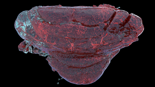
Fluorescent techniques can help scientists see structures such as the vasculature of the brain. Image courtesy of Daniel Stabley, PhD.
How does fluorescence microscopy help St. Jude scientists?
Imagine you are trying to repair a complex machine — but you don’t know what it is; its origin is unknown. The discovery of treatments for pediatric diseases is much like that. The way that dumping an imagined bin of defective microprocessors onto Einstein’s desk won’t fix them, simple intelligence alone won’t lead to the discovery of a treatment for a disease that is poorly understood. One must first understand the logic, the form, the function, the causes and effects and the very rules that govern the behavior of any given biological property. Only then can it be mastered, controlled and altered for a better outcome. This is what we endeavor to do through basic science. Fluorescence microscopy is one of the best tools available to do so by observing the mechanisms underlying disease processes.
Fluorescence microscopy has allowed modern scientists an unprecedented look into the inner workings of cells by showing us individual components in their native context. Instead of the jumble of protists, plant matter and dirt one would observe through a high school biology classroom microscope, fluorescence microscopes can show us specific facets of cellular function in real-time.
For example, in research published in Nature Communications in 2016 from the St. Jude Neuroimaging laboratory and the laboratory of David Solecki, PhD, St. Jude Department of Developmental Neurobiology, it was possible to watch the interaction of specific proteins as a neuron crawled inward from its birthplace in the developing brain, thereby learning that these proteins are essential for normal brain development. In another study to which these labs contributed, published in Science in 2020, fluorescence microscopy combined with other techniques, such as electron microscopy, were used to study how the nuclei of cells change during development. These examples all relate to one common theme: To intervene and repair the malfunctions in cells that give rise to disease, we must understand them in the first place.
Towards the future: Advanced imaging at St. Jude
Fluorescence microscopy is one of the best examples of how the development of novel tools can completely disrupt and invigorate the landscape of science and transform our fundamental understanding of life itself. However, this technology is imperfect, and there is much to be gained by its improvement. For example, fluorescence images become increasingly distorted as we move deeper into tissue, necessitating the development of techniques such as adaptive optics fluorescence microscopy.
It goes without saying that we must not merely be content with the techniques we have but should also pursue the development of the next generation of tools. Developing new microscopy methods is a complex task that requires diverse teams that span a multitude of expertise, from physics to chemistry to biology, and is best done in close collaboration with the scientists who will use the technology in their own research after it is developed.
St. Jude is an environment where this collaboration comes naturally and has therefore been an ideal place to push the frontiers of modern imaging through various initiatives, including the Neuroimaging Laboratory. Through the Neuroimaging Laboratory, we are expanding access to and competency in advancing imaging methods at St. Jude. It is my hope that the tools we develop today will give biologists unparalleled access to the unseen places that lie at the heart of catastrophic diseases, leading to a brighter future for children everywhere.


