St. Jude Family of Websites
Explore our cutting edge research, world-class patient care, career opportunities and more.
St. Jude Children's Research Hospital Home

- Fundraising
St. Jude Family of Websites
Explore our cutting edge research, world-class patient care, career opportunities and more.
St. Jude Children's Research Hospital Home

- Fundraising
The life and death of cells in early brain development

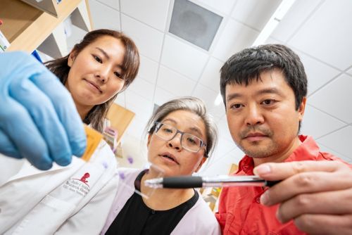
(L to R) First author Yurika Matsui, PhD, corresponding author Jamy Peng, PhD, both of St. Jude Department of Developmental Neurobiology, and co-author Beisi Xu, PhD, of St. Jude Center for Applied Bioinformatics examine samples in the lab.
When fertilized cells begin to divide and develop into an embryo, it is easy to fixate on the concept of growth, of new life. However, life is only one side of the equation when it comes to development. Cell death also plays a role in shaping how tissues, particularly the brain, develop.
Researchers at St. Jude are trying to better understand the role that cell death plays in neurodevelopment. The way fingers and toes develop is a classic example of the importance of cell death in developmental biology.
“Our limb digits, like fingers and toes, are formed by actively going through cell death to make the proper structure,” said Yurika Matsui, PhD, a postdoctoral researcher in the lab of Jamy Peng, PhD, St. Jude Department of Developmental Neurobiology.
You can think of this phenomenon as a sculptor chipping away the excess stone to reveal the figure inside: our fingers, toes, and most other parts of our bodies are formed by the precise control of life and death during early development. Matsui and Peng wanted to know more about the role of cell death in the formation of the brain.
Uncertainty behind neural progenitor cell fate
The brain is formed through an astonishingly well-controlled process. This process is driven by neural progenitor cells (NPCs), the foundation of the brain, and their perfectly timed division, death and differentiation. While multipotent stem cells can develop into many adult cells, NPCs are slightly different because they can only develop into neural cells. Their development and differentiation rely on Polycomb repressive complex 2 (PRC2). This protein complex maintains genetic programs that direct cells to grow into the tissues that make up the brain. “PRC2 is important because its mutations and dysfunction lead to a wide range of pediatric cancer types,” explained Peng.
But what occurs when NPCs die during early brain development? “We in the developmental field didn’t really know what the molecular mechanism of death was in those cells,” explained Matsui.
Published recently in Nature Communications, corresponding author Peng and first author Matsui uncovered the culprit responsible for regulating NPC death and survival to ensure the correct shape, size and formation of the brain.

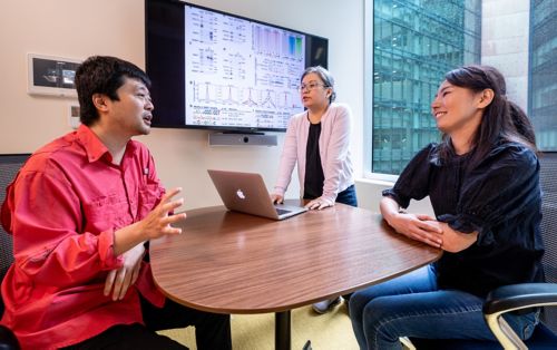
(L to R) Co-author Beisi Xu, PhD, of St. Jude Center for Applied Bioinformatics, corresponding author Jamy Peng, PhD, and first author Yurika Matsui, PhD, both of St. Jude Department of Developmental Neurobiology discuss their bioinformatics data.
Examining the motive to find the suspect
Knowing that the pathway responsible for cell fates was tightly regulated, Peng began by looking closer at PRC2. “To understand the fundamental regulation of cell differentiation by PRC2, we underwent a proteomics approach,” Peng explained. This approach was to pinpoint any protein interactions that were taking place. “We identified a protein called Smad nuclear interacting protein 1 (SNIP1) as a binder of PRC2 in the NPCs,” Peng continued.
SNIP1 is part of a long cascade of proteins that regulate cell development and growth, but its ability to promote cell division also means it can regulate tumor growth. Additionally, mutations in SNIP1 have been shown to lead to neurodevelopmental disorders and epilepsy.
Working with Beisi Xu, PhD, St. Jude Center for Applied Bioinformatics, they interrogated the importance of SNIP1 in NPCs using mouse models of early brain development. They found that SNIP1 and PRC2 work together like the two ends of a seesaw to regulate cell survival and death. “We found that together, PRC2 and SNIP1 regulate gene expression positively and negatively to facilitate cell death, neural differentiation and the self-renewal of NPCs,” Matsui explained. “SNIP1 and PRC2 essentially function as a regulatory system to prevent cell death,” Peng added.
Peng, Matsui and Xu are now looking at how they can build off this discovery. Of note is the role that SNIP1 plays in other cells. “SNIP1 has other molecular roles, such as RNA processing and supporting oncogene activities,” Matsui said. “I want to see if these other roles cooperate to give rise to the phenotypes we saw in the SNIP1-depleted cells.”
Peng added, “I think this particular protein is highly promising for future impactful discoveries.”


