St. Jude Family of Websites
Explore our cutting edge research, world-class patient care, career opportunities and more.
St. Jude Children's Research Hospital Home

- Fundraising
St. Jude Family of Websites
Explore our cutting edge research, world-class patient care, career opportunities and more.
St. Jude Children's Research Hospital Home

- Fundraising
Preventing neurodevelopmental disorders through genetic compensation - Progress

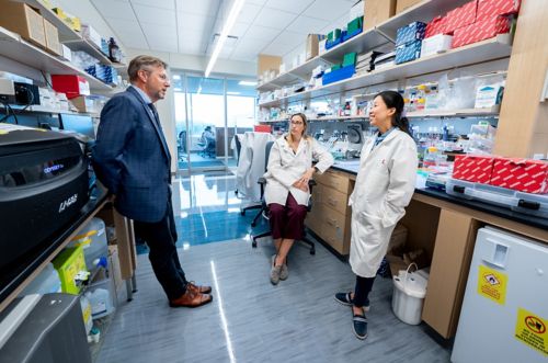
Co-corresponding author J. Paul Taylor, MD, PhD, first author Ane Korff, PhD, and co-corresponding author Hong Joo Kim, PhD, of St. Jude Department of Cell & Molecular Biology.
It would be much easier to treat disease if there was always one pathway, protein, or gene that could be pointed to as the root cause and correcting that problem would provide a direct route to treatment. However, the situation is usually not that simple. The complex, interwoven nature of our biochemical landscape means that by design, our body has checks and balances. A misaligned system can often be rescued by a related protein or the up- or down-regulation of alternative pathways. To find the chain of events that causes disease, the layers of effect must be peeled back, piece by piece. Only by shining light on this complexity can researchers tackle the root pathogenicity.
In 2016, the HNRNPH2 gene was flagged for the first time in individuals showing signs of intellectual disability, developmental delay and autism spectrum disorder. The HNRNPH2 gene codes for a specific protein within a group of proteins called the heterogenous nuclear ribonucleoprotein (hnRNP) family. These hnRNP proteins are pivotal to many processes tied to RNA handling, including transcription and translation.
“Because of our history working with the hnRNP family, when the mutations in HNRNPH2 were first identified, we were recruited to address the role the protein plays in this disorder,” said Hong Joo Kim, PhD, St. Jude Department of Cell & Molecular Biology. Kim works in the laboratory of J. Paul Taylor, MD, PhD, St. Jude Scientific Director. Over time, HNRNPH2 mutations have been found in over 30 individuals with traits of neurodevelopmental disorders.
Published in The Journal of Clinical Investigation, co-corresponding authors Kim, Taylor and their team, including first author Ane Korff, PhD, shed light on the mechanisms behind this pathogenicity. The work provides an important lens into the biology of this poorly understood disorder, with the potential to develop future therapeutics that target this and related mechanisms.
Mixed signals at the root of disorder symptoms
HNRNPH2 is a gene that is found exclusively on the X chromosome. Women have two copies of the chromosome (XX), while men have only one (XY). Mutated genes on the X chromosome can seriously affect the body, including intellectual disability and developmental delay in the case of HNRNPH2. However, because women have a second version of the gene that functions normally, the effect can be partially mitigated. This is not the case in men, where the effects of a mutation on the X chromosome can be more disastrous.
“The first identified patients with HNRNPH2 mutations were all female patients,” explained Kim. “For males, who have a single X chromosome, the effect of mutation is so severe compared to females that the number who survive after birth is very low.”
The researchers found that the HNRNPH2 mutations affect the protein’s nuclear localization signal (NLS). The NLS acts as a protein’s security clearance to enter and exit the nucleus. Considering that the function of hnRNPH2 is vital to shaping RNA, mutations affecting the NLS can be disastrous, resulting in the protein losing security access and stranding the protein outside the nucleus. From this point, how the protein contributes to neurodegeneration has been a mystery that Kim and Taylor are looking to address.
When they looked at the disease form of the protein with a mutated NLS, the researchers unsurprisingly saw that access to the nucleus had been reduced. “There was about a quarter of the total protein that was still in the cytoplasm,” Kim explained. But this alone did not indicate the root cause of the disorder. “In terms of disease mechanism, is its reduced presence in the nucleus causing a loss of a function,” Kim questioned, “or is the protein accumulation in the cytoplasm causing a newly gained function?”
Absence is much better than mutation for hnRNPH2
The researchers arduously designed mouse models and human cell lines, crucial components to get to the bottom of this mystery. The cells and mice were either completely lacking HNRNPH2 or contained the disease-causing mutation, referred to as a “knock-out” and a “knock-in,” respectively. “If our knock-in mice behave similarly to the knock-out mice, then this would suggest that the mutation causes a loss of function,” Kim explained. “If not, then the mutation might be causing a gain of a function.”
Based on preliminary data, all signs pointed to a loss-of-function pathway, a disease effect brought about by hnRNPH2 having reduced access to the nucleus. However, further probing turned that idea inside out.
“To our surprise, our knock-out mice were completely normal,” exclaimed Kim. “They barely had any differences compared to the normal mice.” Meanwhile, the knock-in mice with the mutated protein showed signs of the disorder, demonstrating not only that the researchers successfully created a faithful disease model but also that this was not a simple loss-of-function story.
Considering the validation of their cell lines, the researchers next looked at the expression patterns of other hnRNP family members to see if there was any correlation. One protein stood out. “When we generated the knock-out lines,” Kim said, “we saw in both the human cells and in mouse lines that hnRNPH1 levels go up.”
For disease mitigation, family is everything
As the name suggests, hnRNPH1 is a close relation to hnRNPH2. The main difference lies in their expression patterns over time. H1 is predominantly found in the developmental life stages before dropping off. H2 levels are steadily maintained throughout life at low levels. This is due to the body’s different requirements during development and our subsequent lifetime. “We think that H1 and H2 are functionally redundant, and H1 is more important before birth during embryo formation,” Kim stated, “whereas H2 has a very consistent role throughout the lifespan.”
However, the researchers found that H1 levels were elevated in the knock-out mice. These results implied that H1 expression levels were raised to compensate for the complete loss of H2, a fail-safe to rescue the cells.
And what of the disease mutant? “The situation becomes totally different because H2 protein is still going to be expressed, but it’s not really functional,” explained Kim. “Because there is still protein, the compensatory role of H1 does not kick in.”
This effect could even be tuned. When the researchers slowly reduced levels of H2, they found that H1 levels were initially not bothered. But at a certain point, Kim explains, “At around 80% reduction of H2, we see the H1 upregulation, and the level becomes over 150%.” This more than makes up for the loss of H2.
Importantly, these fundamental genetic insights also identified a strategy for therapeutic intervention using antisense oligonucleotides (ASOs), synthetic RNA designed to bind to naturally occurring messenger RNA, a treatment strategy which has found success in the treatment of spinal muscular atrophy (SMA) among other neurodevelopmental disorders. In the case of hnRNPH2-related disorder, the compensatory relationship between H1 and H2 suggests that one could design an ASO to decrease the levels of H2.
“Since our studies showed that a complete knock-out of H2 in cells and mice did not cause any detectable ill effects, lowering the levels of H2 using an ASO would be predicted to be well tolerated in patients,” said Korff. “But more importantly, knocking down H2 to very low levels would be predicted to trigger compensatory upregulation of H1, with potential therapeutic benefits.”
Taylor was quick to highlight how the discovery of the H1-H2 relationship and the mechanistic insights from the mouse models can be carried forward. “The findings from this study represent crucial documentation on the path to introducing a therapy for children with mutations in HNRNPH2,” he said. “It’s an essential pre-clinical step.” Indeed, the design of ASO-based treatment for hnRNPH2-related disorder is now moving forward as part of the Blue-Sky project called GEMINI (Genomic Medicines Initiative), a program launched this summer within the Pediatric Translational Neuroscience Initiative.
The invaluable insight has pushed the boundary of what researchers know and understand about this neurodevelopmental disorder characterized by a mutation in HNRNPH2. The work also demonstrates that disease pathogenesis is not a simple “one-shot” mechanism. Instead, it is a balance between H2 mutation causing a loss of function and impaired compensation by H1, or perhaps some mechanism of gain-of-function in the cytoplasm. The work has revealed how complex relationships can lie at the root of dysfunction. However, as these findings prove, the models designed for this work are the real gift. The combinative effect they have will allow for a new paradigm to be reached in future research into hnRNP-linked neurodegenerative disease.


