St. Jude Family of Websites
Explore our cutting edge research, world-class patient care, career opportunities and more.
St. Jude Children's Research Hospital Home

- Fundraising
St. Jude Family of Websites
Explore our cutting edge research, world-class patient care, career opportunities and more.
St. Jude Children's Research Hospital Home

- Fundraising
Clinical Practice Project
Standardizing imaging of the joints for assessment of AVN
About the center
In an effort to standardize imaging of the joints for assessment of AVN with the St. Jude Affiliates, we are providing our MRI AVN Siemens Scanning Protocols, representative images of each joint, and tips for imaging and interpreting these studies. We also compiled the current literature regarding AVN in pediatric oncology patients for your reference. Do not hesitate to contact us if you have questions or need clarification.
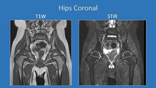
St. Jude routine scanning protocols include:
- T1W coronal
- STIR coronal
- Gradient echo sagittal through the joint
- We use an 18 channel coil for all joints except the ankles, for which we use a head coil.
- We do NOT routinely do axials or post-contrast imaging for any joint.
- For evaluating the hips, knees and ankles, the right and left joints are imaged simultaneously within one large field of view rather than separately with smaller FOVs. This has several benefits: 1) it shortens table time and 2) facilitates the radiologist's comparison to prior exams.
- For shoulders and elbows, the right and left joints are imaged separately using a relatively smaller FOV. This approach avoids artifact from breathing motion and reduced image quality due to large FOV that spans the entire chest.
- Key features to assess on the imaging are: 1) whether foci of AVN are subarticular in location and 2) whether the articular surface cartilage is smooth or irregular and intact or flattened.
AVN MRI representative images of each joint
AVN files for download
These files can be viewed using a DICOM viewer or by adding the images to PACS. For questions about image viewing, please contact Nancy Foster at nancy.foster@stjude.org.
Current literature
- Osteonecrosis in children after therapy for malignancy
- Unique MRI findings as an early predictor of osteonecrosis in pediatric acute lymphoblastic leukemia
- Utility of early screening magnetic resonance imaging for extensive hip osteonecrosis in pediatric patients treated with glucocorticoids
- Osteonecrosis of the Shoulders in Pediatric Patients Treated for Leukemia or Lymphoma: Single-Institutional Experience
