St. Jude Family of Websites
Explore our cutting edge research, world-class patient care, career opportunities and more.
St. Jude Children's Research Hospital Home

- Fundraising
St. Jude Family of Websites
Explore our cutting edge research, world-class patient care, career opportunities and more.
St. Jude Children's Research Hospital Home
- Fundraising
Zachary Abramson, MD, DMD
Developing computer-aided 3D models to assist in the diagnosis and treatment of pediatric tumors
Overview
3D modeling is a useful tool for medical providers, making substantial impact in fields such as dentistry and cardiology. However, its application to pediatric cancers has been limited due to difficulties in imaging soft tissue tumors in this patient population. My work aims to develop and optimize the process of building 3D models to aid in the diagnosis and treatment of pediatric cancers.
In the news
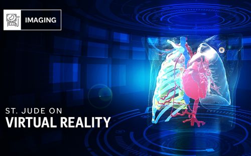
Seeing is believing: Bringing virtual reality into the clinic
See how St. Jude investigators are using virtual reality in their work.
Abramson Research Summary
Radiology has historically been a valuable asset for diagnostic medicine. With advances in imaging technology, it is playing a more central role in all aspects of treatment strategies. This is especially true for the field of pediatric radiology, where oncologic imaging is being called upon to assist in pre- and intra-operative planning.
My work focuses on the development and optimization of computer-aided 3D modeling to assist in the diagnosis and treatment of pediatric tumors.
3D modeling for pediatric tumors
The treatment strategy for children with soft tissue tumors, such as high-risk neuroblastoma and bilateral Wilms tumors, often involves surgical resection of the tumor. Resection of these tumors can be complex for many reasons, including vascular encasement. Therefore, it is imperative to have an accurate understanding of the relationship between the tumor and the architecture of the surrounding area in order to have the most successful surgical outcomes.
3D models have become an important part of the surgical planning process, one that stands to impact surgical outcomes and, furthermore, patient outcomes. To make an accurate 3D model, we must have pristine images as the data from these images are the foundation for the model and, therefore, must be accurate, precise and contain scant imaging noise. As an expert in pediatric oncologic imaging, I am developing and optimizing the imaging protocols that inform the building of 3D models.
We have developed a split-injection contrast-enhanced computed tomography (CT) technique for cases of neuroblastoma, allowing us to obtain all necessary images from a single scan. This concurrent imaging approach lessens a patient’s exposure to radiation and allows us to bypass the amalgamation of multiple acquisitions, which has been known to introduce error in the 3D model.

We work closely with faculty in the Department of Surgery to be able to utilize our 3D models in the most impactful ways. Canonically, these 3D models were visualized on a 2D display, which led to a phenomenon called size-distance uncertainty. Surgeons were not always able to accurately grasp the size and location of tumors because of limitations based on a 2D display.
By translating the 3D model into a 3D display via virtual reality headsets, we have added stereoscopic vision and parallax to resolve the issue of size-distance uncertainty. I work with image processing specialists to translate the 3D model into a virtual existence so surgeons can use it during pre- and intra-operative planning.
In addition, this technology is also being used by surgeons during consultations with patients and family. Surgeons and family can wear virtual reality headsets and occupy the same virtual space when discussing surgical options. This approach elevates the ability of the treatment team to inform patients and family ultimately leading to better care.
3D visualization as an emerging field
I am interested in exploring the impact of utilizing 3D visualization for all parties involved – from the perspective of surgeons, patients and family members. Furthermore, I am working to educate industry on the benefits and necessity of 3D technology for medical imaging. Industry’s input, along with guidance from advanced practice providers in radiology, will advance the capabilities of 3D imaging and allow us to provide superior patient care.
Learn more
Selected Publications
About Zachary Abramson
Dr. Abramson is a pediatric radiologist with a specialization in oncologic imaging. He holds a Bachelor of Science degree from the University of Michigan, a Doctor of Dentistry degree from Harvard School of Dental Medicine and a Medical Degree from the University of Tennessee College of Medicine. He has trained as an intern at Massachusetts General Hospital (oral and maxillofacial surgery) and the University of Tennessee College of Medicine (internal medicine). Furthermore, he completed a radiology residency program at Baptist Memorial Hospital in Memphis, TN, and a fellowship in pediatric radiology at Le Bonheur Children’s Hospital in Memphis, TN.
Dr. Abramson is board certified in Radiology with subspecialty certification in Pediatric Radiology and serves as an Assistant Member in the Department of Radiology. His unique educational background and skillset allow him to develop and optimize 3D modeling for surgical planning. He is also passionate about education pertaining to the capabilities of 3D visualization as well as expanding the scope and reach of this type of imaging.

Contact us
Zachary Abramson, MD, DMD
Assistant Member, St. Jude Faculty
Department of Radiology
MS 220, Room I3120
St. Jude Children's Research Hospital
Memphis, TN, 38105-3678 USA GET DIRECTIONS