St. Jude Family of Websites
Explore our cutting edge research, world-class patient care, career opportunities and more.
St. Jude Children's Research Hospital Home

- Fundraising
St. Jude Family of Websites
Explore our cutting edge research, world-class patient care, career opportunities and more.
St. Jude Children's Research Hospital Home

- Fundraising
Comparative Pathology Core (CPC)
The Comparative Pathology Core Laboratory (CPCL) provides a wide range of services with the mission to aid investigators using preclinical models.
OVERVIEW
The Comparative Pathology Core Laboratory (CPCL) provides a wide range of services with the mission to aid investigators using preclinical models. Our major areas of service include clinical diagnostic, necropsy, and histology laboratories and research support from four board-certified veterinary pathologists.
- Clinical Diagnostic Laboratory: Hematology, serology, bacteriology, parasitology, coagulation panels, blood chemistries (standard plus specialized assays such as insulin, leptin, bile acid, alpha-fetoprotein, testosterone, estrogen, and FSH), mouse sperm analysis, and molecular diagnostics (PCR)
- Necropsy Laboratory: Full necropsy service, whole body perfusion, tissue collection, training investigators in necropsy and tissue collection techniques
- Histology Laboratory: Full range of histology services, special histochemistry stains (~30 stains), immunohistochemistry (~300 assays), In situ hybridization, multiplex labeling, frozen sections, custom IHC/ISH assay development, whole slide scanning
- Pathologists: Histopathology evaluations, consultation on model development, image and immunohistochemistry analysis support, laser capture microdissection


IMPACT
The Comparative Pathology Core Laboratory (CPCL) at St. Jude provides expert, readily available and affordable research pathology support. Our goal is to support interdisciplinary research that uses animal models to study the pathogenesis of pediatric cancers as well as metabolic and infectious diseases, in evaluating the efficacy and toxicity of experimental therapeutic compounds, and in determining the functions of specific genes and gene products (transgenic and knockout mouse technology). The CPCL staff includes four ACVP board-certified veterinary pathologists with extensive experience in comparative pathology, and certified histology and clinical laboratory technicians. State-of-the-art facilities support complete necropsy, histology, and immunohistochemistry services and include a fully equipped clinical pathology laboratory that provides hematology, clinical chemistry, parasitology, molecular diagnostics, and microbiology testing services.
Equipment
Diagnostic pathology
- Forma dual stack CO2 incubator
- BD Bactec 9050 blood culture syste
- Vitek compact bacteriology identification system
- Whitley DG250 Anaerobic workstation
- Wescor stainer/cytospin centrifuge
- Beckman refrigerated centrifuge
- Eppendorf high speed tabletop centrifuge
- Two Forcyte Hematology analyzers with impedance and laser technology
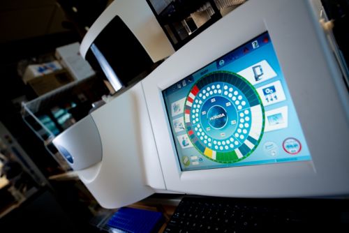
- Horiba P400 Chemistry analyzer
- Hamilton Thorne IVOS sperm analysis instrument
- IDEXX VetLab UA instrument (urinalysis)
- Premiere Slide warmer XM-2002
- Fisherbrand Isotemp dry bath
- Siemens BFTII coagulase instrument
- Biotek ELx800 plate reader
- Fisher scientific accuWash plate washer
- Two Qiagen Qiacubes for automated nucleic acid extraction using Qiagen spin kits
- Qiagen Qiagility automated fluid handling system for PCR setup
- (2) Cepheid Smart Cyclers for real-time PCR
- ABI StepOnePlus real-time PCR system
- Omni International Bead Ruptor homogenizer
- Thermo Precision convection laboratory incubator
- Eppendorf Thermomixer R
- Forma Ultra low freezer
- Kenmore compact refrigerator
- Three fisher scientific compact freezers
- IsoTemp glass door refrigerator
- Fisherbrand Isotemp sliding door refrigerator
- Kenmore standard stainless refrigerator/freezer
- Tuttnauer tabletop autoclave
- Two Olympus BX41 multi-head teaching microscopes
- Zeiss Stemi 2000 dissection scope
- Biotek EPOCH plate reader
Necropsy/Histology
- Excelsior ES Tissue Processor ( Thermo Fisher)
- Excelsior AS Tissue Processor ( Thermo Fisher)
- 2 HistoStar Embedding Centers (Thermo Fisher)
- 4 HM 355S Microtomes (Thermo Fisher)
- HistoCore Autocut Microtome (Leica)
- Gemini Automated Routine Stainer (Thermo Fisher)
- Dako CoverStainer (Agilent), CM 3050S Cryostat (Leica)
- BondMax IHC Stainer (Leica)
- Bond RX IHC Stainer (Leica)
- 2 Ventana Discovery XT IHC Stainers (Roche Medical)
- IntelliPath Automated IHC Stainer (Biocare Medical)
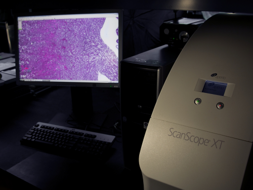
- Dako Artisan Special Stainer (Agilent), Aperio ScanScope XT (Leica)
- Arcturis XT laser microdissection system (Thermo Fisher)
- PrintMate AS Cassette Printer
- 6 Downdraft dissection work stations (Thermo Fisher)
- Downdraft necropsy/dissection table (Thermo Fisher)
- Olympus dissecting microscope with digital camera system, 4 research-grade microscopes with digital camera systems
- Nikon multihead teaching microscope
- Arcos Block Management System (Thermo Fisher)
- Arcos SL Slide Management System (Thermo Fisher)
Services
CPCL Necropsy Service
- Gross examination and sample collection in various laboratory animal species
- Interpretation of findings and preparation of reports
- Consultation with research personnel as needed
- Comprehensive macroscopic and microscopic examinations
- Emphasis on phenotypic characterization of genetically engineered mice
- Use of downdraft tables, whole-body perfusion fixation pumps, and camera setups
- Experienced necropsy personnel for various animal species
CPCL Histology Laboratory
- Routine and advanced histology services
- Custom immunohistochemistry services
- Preparation and staining of paraffin-embedded and frozen sections
- Standard and advanced histochemistry and immunohistochemistry
- mmunohistochemical assay development
- Laser capture microdissection
- Whole-slide scanning and image analysis
- Expert pathologists for interpretation of morphological changes and lesions
- Knowledge of normal anatomy and physiology in various animal models
- Assistance in selecting appropriate animal model systems and experimental pathology assays
- Support for model development, validation, experimental design, sample collection, fixation, and data interpretation
- Consultation and training with multiheaded microscopy and photomicrographic equipment

[Collaboration] is at the heart of our Comparative Pathology Core. Our pathologists work to support scientists with different animal models, as interpreting histopathology is crucial to their research efforts.
David Ellison, Department Chair
CPC by the numbers
3,500
3,500 necropsies performed annually
CPC by the numbers
100K
100,000 H&E stained slides annually
CPC by the numbers
25K
25,000 IHC slides annually
CPC by the numbers
200+
More than 200 authorships on peer-reviewed publications
CPC by the numbers
50+
More than 50 investigator partnerships
Director
-
View Details
Laura Janke, DVM, PhD, DACVP
Member, St. Jude Faculty
Director, Comparative Pathology Core
Email
laura.janke@stjude.orgLaura Janke, DVM, PhD, DACVP
Member, St. Jude Faculty
Director, Comparative Pathology Core
Affiliations
Research Interests
- Phenotyping of genetically engineered mice
- Assessment of treatment efficacy and toxicity in mouse models of pediatric cancers
- Use of immunohistochemistry to aid in the investigation of tissue- and cell-specific molecular changes
- Use of histomorphometry and whole slide imaging to assist in the generation of objective data regarding histopathological lesions or antigen expression
Contact Information
Laura Janke, DVM, PhD, DACVP
Pathology
MS 250, Room C5036
St. Jude Children's Research Hospital
262 Danny Thomas Place
Memphis, TN 38105-3678

